Experiments
with the modularEEG
EEG-Feedback
Here are some infos about an EEG-Feedback session(from 2002-10-27) with
the
modularEEG.Following session parameters were used:
- audio feedback
- filter center freq=9.4Hz, bw=2Hz
- feedback threshold=30uVp-p
- electrode positions:
- CH0: Cz-Au2
- DRL: on lower right leg
bwview raw/256:s sessionname.pcm) written by Jim Peters. Simply use the following command line: A description of the events during ON-OFF-ON training marked in the data:
021027alpha fb9.4Hz th=30uVp-p ON-OFF-ON_EEGtxt.txt
BTW: The skin under the electrodes (Cz,Au2,pasted with 10/20 wax, DRL electrode w/o) was unprepared. Due to moisture buildup during the session, the electrode impedance probably decreased. As a result the mains hum (50Hz spectral line) decreased over time.
Some screenshots of the session from bwview:
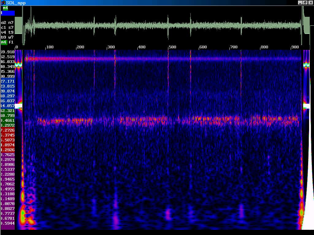
021027alpha fb9.4Hz th=30uVp-p ON-OFF-ON_EEG_M4.png
Same session, display mode m6:
021027alpha fb9.4Hz th=30uVp-p ON-OFF-ON_EEG_M6.png
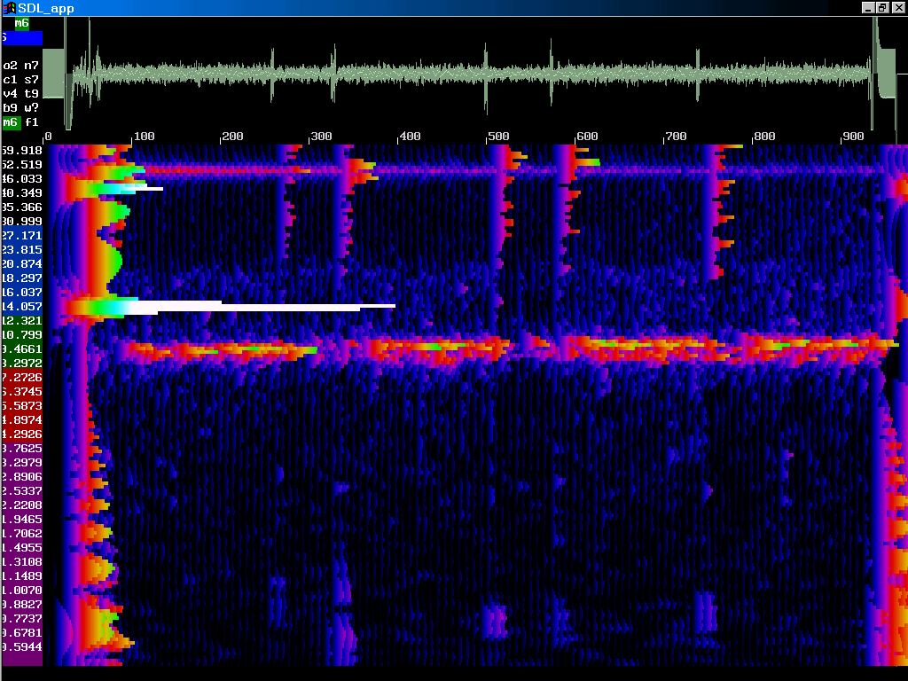
ECG with the modularEEG
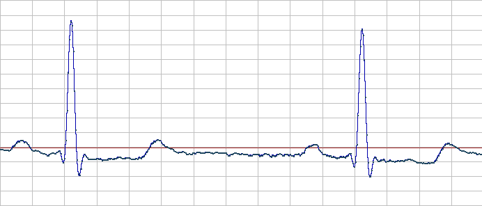
(2002-11-02)ECG has a R-spike amplitude of about 1mV. Because the strong asymmetry of the signal, a total range of at least 2mVp-p for fullscale is necessary. To have headroom for movement artifacts etc. 4mVp-p would be desirable.
So in order to record ECGs with the modularEEG, the gain has to be lowered. This can be easily done by replacing e.g R214 and R215 (for channel2, modEEGamp_v0.05) with 100k. The gain of the first stage is thereby lowered from 12.2 to about 1.(BTW: This is not the best way to lower the total gain, because the signal to noise ratio of the INA114 is better when using higher gains).
The current version modEEGamp_v1.1.0 (2003-11-01) does not need the above modifications. Simply adjust P202 and P203 to the lowest available amplification and you can record ECGs.
(picture: ECG voltage over time)
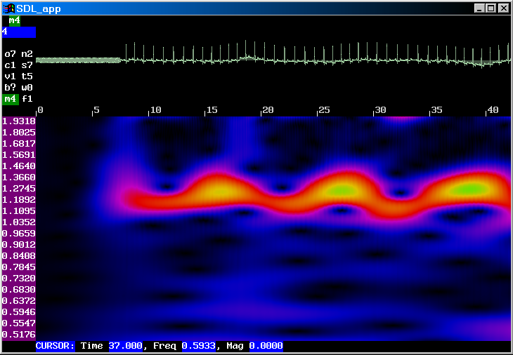
The recorded ECG data can be viewed with bwview (http://uazu.net/bwview/) also. Simply use the following command line:
bwview raw/256:s sessionname.pcm
In the the joint time-frequency-display (JTF) a periodic modulation of the heart rate from ca. 1.1Hz to 1.3Hz can be seen. This is due to respiratory sinus arrhythmia (RSA).
Respiratory Sinus Arrhythmia
In healthy hearts, heart rate varies across the breathing cycle. Heart rate increases during inspiration and decreases with expiration. This pattern observed in normal hearts, is misleadingly called the respiratory sinus arrhythmia (RSA).Cited from http://www2.truman.edu/shaffer/cardio_2001.htm (a part of the Biofeedback bootcamp):
RSA depends on parasympathetic (vagus nerve) regulation of the heart. While disruption of vagal signals to the heart almost completely ends RSA, interference with sympathetic signals does not affect it (Papillo & Shapiro, 1990).
The RSA indexes the heart’s adaptability to changing demands. Ideally, a patient will show a large-amplitude RSA that is perfectly in-phase with respiration. The RSA may be reduced or absent in thoracic breathers and paradoxically when breathing is too slow. The absence of an RSA may be a strong risk factor for heart attack.
(The above URL is invalid now, but mirrored at http://www.psychosensory.com/files/bootcamphome_2001.zip, 6.5mb)